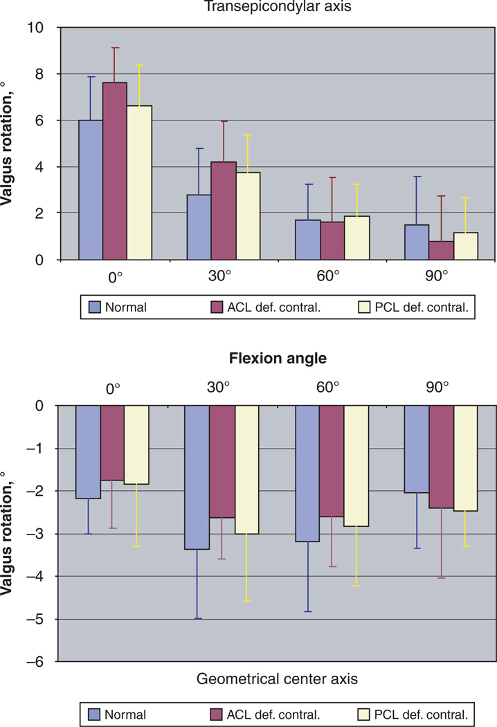
Figure 5.
Varus-valgus alignment of the tibia relative to the femur in subjects with bilateral healthy knees (normal) and patients with contralateral ACL (ACL def. contral.) and PCL (PCL def. contral.) deficiency. Kinematics were analyzed using the transepicondylar femoral axis-based (top) and geometric center axis-based (bottom) coordinate systems. Positive values denote a valgus orientation of the tibia, while negative values represent a varus tibial orientation. No significant differences between the groups were detected at all flexion angles (P > .95).

