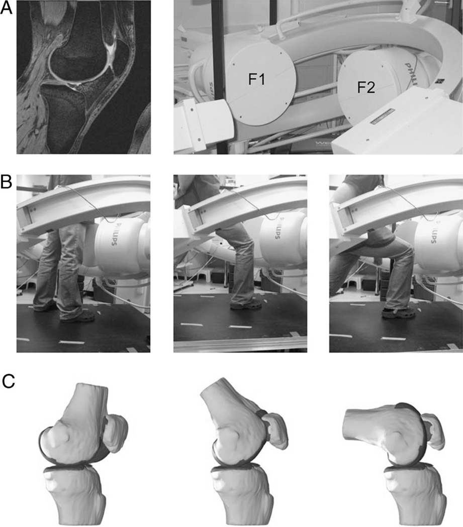
FIGURE 1.
A, The combined MR and dual fluoroscopic imaging technique, in which 3D knee models are created from a series of sagittal MR images (image left), and the motion of the patient’s tested knee is recorded using two orthogonally placed fluoroscopes (image right). B, In the present study, the tested activity was a single-leg quasi-static lunge at 0°, 30°, 60°, 75°, 90°, 105°, and 120° of flexion while the upper body remained upright (only three flexion angles are shown for illustrative purposes). C, The 3D meshed knee models and the series of dual fluoroscopic images were combined to reproduce the knee positions. F1, fluoroscope 1; F2, fluoroscope 2.

