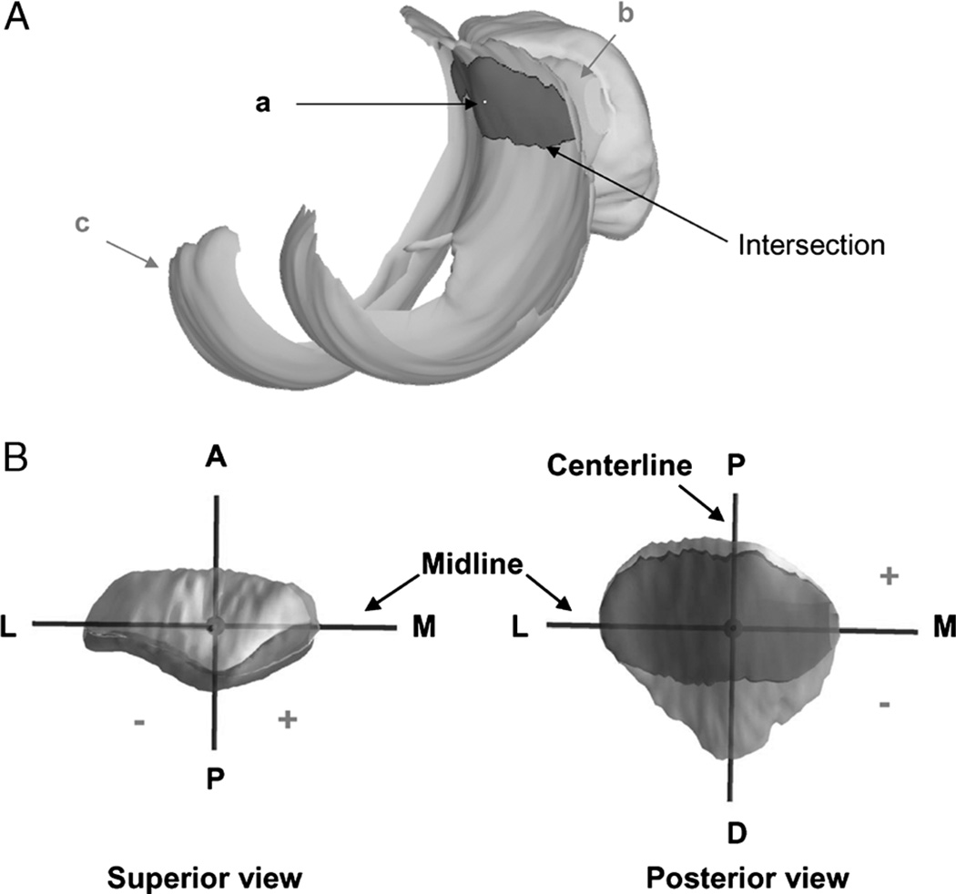
FIGURE 4.
A, The centroid (a) of the intersection of the patellar (b) and femoral (c) cartilage was used to determine the patellofemoral contact locations. B, The coordinate system on the patellar cartilage surface for patellofemoral cartilage contact analysis. The proximal (P)–distal (D) axis was called the centerline. The medial (M)–lateral (L) axis was called the midline. Contact proximal to the midline and medial to the centerline was positive. Reprinted with permission from Van de Velde et al. Am J Sports Med. 2008 Jun;36(6):1150–9.

