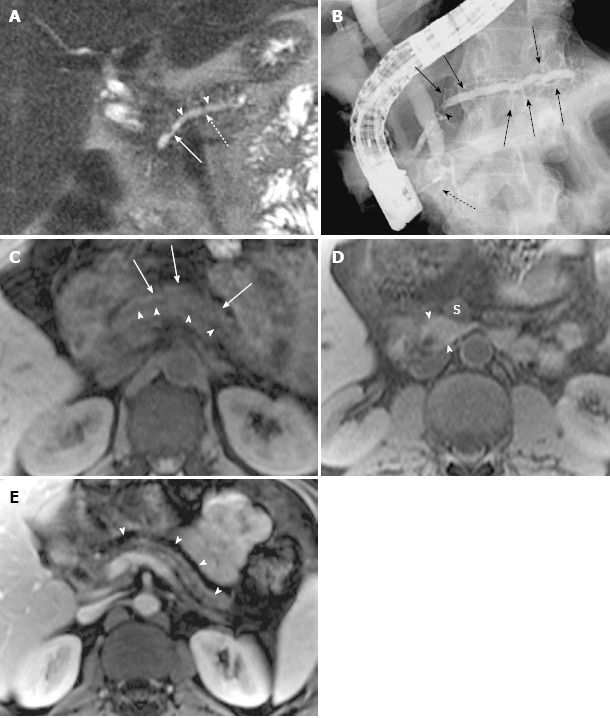
Figure 5.

Magnetic resonance cholangiopancreatography and magnetic resonance imaging of recurrent acute pancreatitis only involving the dorsal pancreas, with endoscopic retrograde cholangiopancreatography correlation, in a 42-year-old man with several episodes of abdominal pain. A: Coronal-oblique, thin-section half-Fourier rapid acquisition with relaxation enhancement magnetic resonance (RARE-MR) cholangiogram [infinite/95 (effective), 3-mm section thickness] shows remarkable dilatation of the dorsal pancreatic duct (dotted arrow) with a conspicuous stricture (solid arrow) and severe side branch ectasia (arrowheads); B: Endoscopic retrograde cholangiopancreatography shows the dilatation of dorsal pancreatic duct and duct of Santorini with a well-seen stricture (arrowhead) and remarkable side branch ectasia (arrows) continual and proximal to the minor papilla. The intrapancreatic segment of common bile duct (C) is very narrow while the other part of the CBD is dilated. The ventral duct is normal in size (dotted arrow); C: Axial precontrast T1WI SPGR shows a marked decrease of signal intensity of dorsal pancreas (arrows) with ductal dilatation (arrowheads); D: Axial precontrast T1WI SPGR shows that the uncinate of pancreas is normal in size and signal intensity (arrowheads) is much higher than the superior mesenteric vein (S) and similar to the liver; E: Axial postcontrast T1WI SPGR shows the atrophy of dorsal pancreas with delayed enhancement (arrowheads) and dilatation of the allied pancreatic duct.
