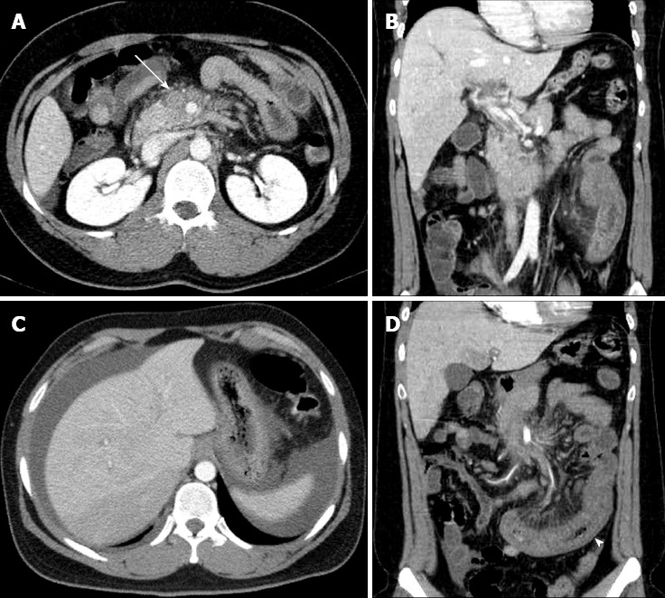
Figure 1.

Abdominal computed tomography demonstrates an acute mesenteric venous thrombosis at the time of initial presentation. A: A thrombus (arrow) and perivenous infiltration at the proximal superior mesenteric vein; B: extension into the portal vein; C: An abnormal fluid collection around the liver and spleen; D: The affected small bowel (arrow head) with long-segment wall thickening and decreased enhancement.
