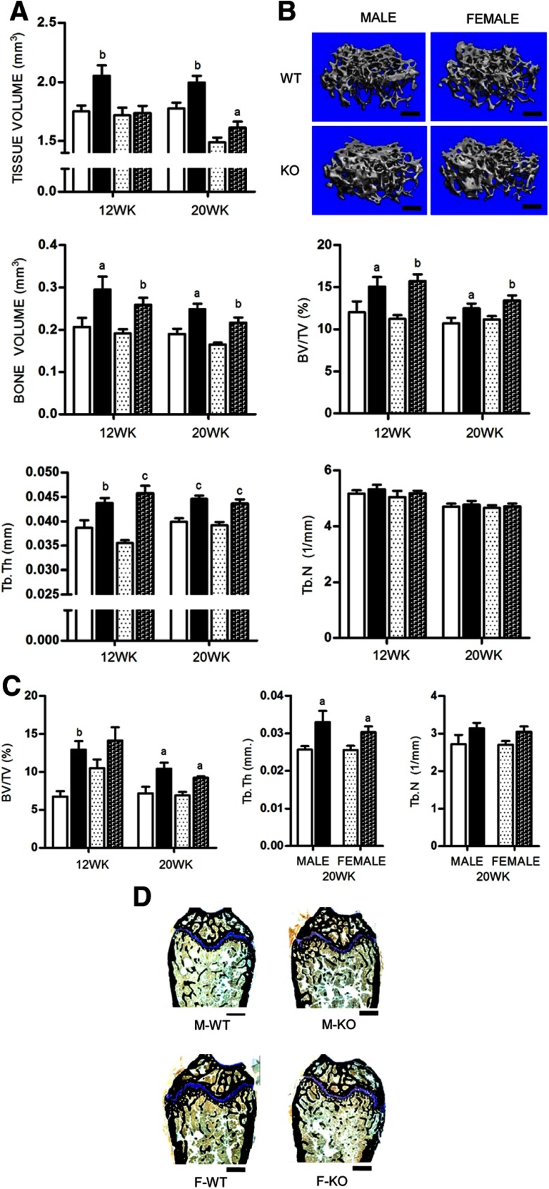
Figure 2.
A, Cancellous bone was assessed by μCT at the distal femurs isolated from male and female mice at 12 and 20 weeks (n ≥ 10 per group). B, Three-dimensional reconstruction μCT renderings of distal femurs from 20-week-old APKO mice compared with age- and sex-matched controls. Representative images were selected from animals whose μCT parameters were closest to the mean of their representative group. C, Histomorphometric analysis of 12- and 20-week-old femurs (n = 5 per group). D, Histomorphometric sections of distal femurs of 20-week-old mice stained for VK and Tetrachrome. All data are mean ± SD. Statistical significance ascertained compared with age- and sex-matched controls. aP < .05; bP < .01; cP < .001. Tb.N, trabecular number; Tb.Th, trabecular thickness. KO, apelin knockout.

