Fig. 7.
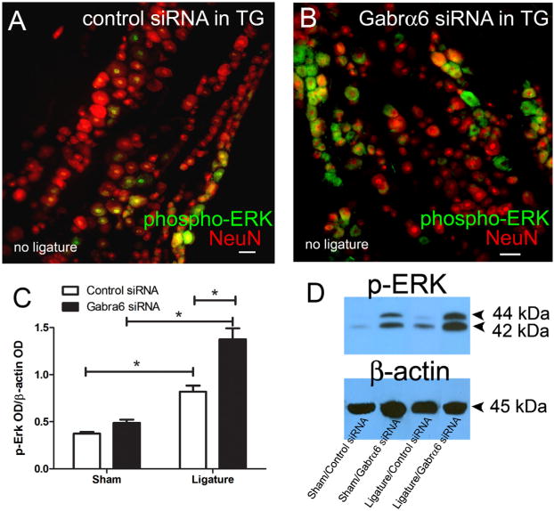
Neuronal activity, as measured by p-ERK, increased after knock-down of Gabrα6. Male rats' trigeminal ganglia were infused with control siRNA or Gabrα6 siRNA 6 days after ligature surgery and 72 h after siRNA infusion the rats were sacrificed and the trigeminal ganglia were isolated. In panels A and B the trigeminal ganglia were stained for p-ERK (green) and NeuN (red). Images are representative of rats in the no ligature groups. Scale bar = 50 μm. (C, D) Western blots were completed using 15 μg of total protein per lane from sham and ligatured rats that were infused with siRNA. Antibodies used in the Western blot were p-ERK and β-actin. (C) Histogram values were reported as a ratio of the optical density of the p-ERK band divided by the optical density of the β-actin band. A two-way ANOVA was performed; independent variables were control siRNA/Gabrα6 siRNA and sham/ligature. The dependent variable was the ratio of the OD. Post-hoc tests were completed comparing each group, *p < 0.05, 10 animals per group. Values are the mean ± SEM. (D) The top right image is a Western blot probed with a p-ERK antibody and this same blot was stripped and probed with a β-actin antibody.

