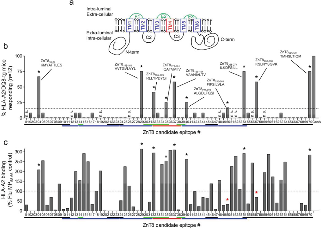
Figure 1. HLA-A2-restricted ZnT8 candidate epitopes.
a ZnT8 protein structure and topology. The 3 intra-luminal loops are shown in green, the 4 extra-luminal domains in black, the 6 transmembrane (TM) domains (TM1–TM6) in blue, except for TM4 (red). b ZnT8 candidate epitopes selected by DNA immunization on HLA-A2/DQ8-transgenic mice. Splenocytes were recalled with the 70 peptides listed in ESM Table 1. Each bar represents the percent of mice responding to the designated peptide or to concanavalin A (ConA) positive control. The dotted line represents the positive cut-off (>15% of mice responding) and selected epitopes are indicated by asterisks. Color lines below X-axis show the position of each epitope within the ZnT8 structure, following the color code of panel a. c Binding of ZnT8 peptides to recombinant HLA-A2.1 in vitro. The same peptide library was tested using the Class I REVEAL binding assay (Proimmune). Results are expressed as percent binding and the dotted line shows the positive cut-off (>100% binding compared to the reference Flu MP58–66 peptide). Asterisks indicate peptides selected by DNA immunization, two of which (red asterisks) are weak binders.

