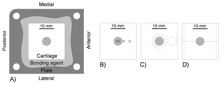
Figure 1.

A) Schematic of osteochondral specimen on base plate, and locations of the B) mechanical tests (circles) and imaging (x’s), C) cuts for biochemistry tests, and D) cuts for histological slides on the osteochondral specimens. The impact area in the center of the specimen is indicated by shading.
