Fig. 1.
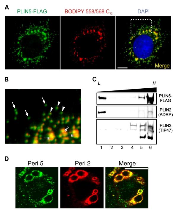
Perilipin 5 localizes to biochemically distinct intracellular pools. A. Confocal images of a representative CHO cell stably expressing the perilipin 5-3X-FLAG construct (green). The perilipin 5-3X-FLAG protein was detected using an anti-FLAG antibody (green). Neutral lipids were stained through incorporation of BODIPY 558/568 C12 (red), and nuclei were stained with DAPI (blue). Scale bar = 10 μm. B. Inset from merged image (right panel in A). Under basal growth conditions, perilipin 5-3X-FLAG localized to both LDs (double arrows) as well as smaller punctate structures in the cytosol. Of the latter, some were associated with neutral lipid staining (arrowheads), while others were apparently devoid of BODIPY 558/568 C12 labeling (arrows). C. Perilipin 5-3X-FLAG-expressing CHO cell lysates prepared following incubation in lipid-loading medium, were subjected to ultracentrifugation using conventional discontinuous sucrose density gradients (0–30%). Immunoblots were prepared using aliquots of equal volume fractions of the gradients, arranged from low (L) to high (H) density, and probed with anti-FLAG, anti-perilipin 2 (ADRP), and anti-perilipin 3 (TIP47) antibodies. Whereas the majority of perilipin 2 and perilipin 3 partitioned into either low or high-density fractions, respectively, perilipin 5-3X-FLAG was recovered in fractions at both extremes of the density gradient. D. Confocal images of a representative CHO cell stably expressing the perilipin 5-3X-FLAG construct (green) and stained with an anti-perilipin 2 antibody (red). Merged images are shown in yellow. Both perilipin 5 and perilipin 2 localize to lipid storage droplets but only perilipin 5 is found on punctate structures.

