Fig. 2.
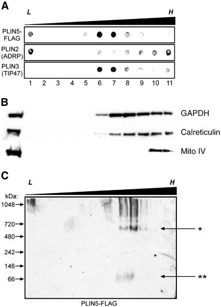
A portion of perilipin 5 is associated with high-density structures of a discrete size. A. Perilipin 5-FLAG-expressing CHO cell lysates were subjected to ultracentrifugation using extended discontinuous sucrose density gradients (0–60%). Dot blots were prepared using aliquots of equal volume fractions of the gradients, arranged from low (L) to high (H) density, and probed with anti-FLAG, anti-perilipin 2 (ADRP), and anti-perilipin 3 (TIP47) antibodies. When the gradients were extended over a wider density range, perilipin 5 was found to partition into both low-density (buoyant) fractions (lane 1) as well as fractions of higher density (lanes 6–7), but was essentially absent in the fractions of highest density (lanes 9–11). B. Marker proteins for cytosol (NADPH), ER (calreticulin) and mitochondria (mito complex IV) are shown. C. Non-denaturing gradient gel electrophoresis was performed using sucrose gradient fractions prepared as described in A. Immunoblots were probed with anti-FLAG antibodies. Immunoreactive material that partitioned into the denser fractions of the gradient also migrated into the gel under non-denaturing conditions, revealing an approximate size of 575 kDa (marked by the asterisk, *). A small amount of immunoreactivity was found migrating near the predicted size of perilipin 5 (double asterisk, **). Please note that each panel contains representative data from different experiments.

