
Fig. 3.
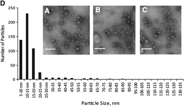
A majority of high-density perilipin 5-containing structures have a size of less than 20 nm. Transmission electron microscopy was performed on material immunoprecipitated from the post-nuclear supernatant isolated from perilipin 5-3X-FLAG-expressing CHO cells grown under basal conditions. A–C Representative images of negative stained immunoprecipitated fractions. Scale bars = 100 nm. D. Histogram showing the size distribution of 500 particles from randomly chosen, non-overlapping microscopic fields, as determined using morphometric analysis.