
Fig. 4.
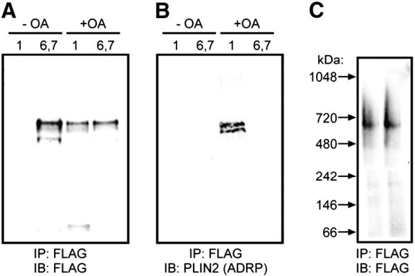
Co-immunoprecipitation of perilipin 5 coated particles confirms a lack of perilipin 2/ADRP in high-density structures. In A–C, cells were either treated with oleic acid (+ OA) or basal growth conditions (− OA). Sucrose gradient (0–60%) ultracentrifugation was performed and fractions 1 (low density) or 6 and 7 (high density) were immunoprecipitated with anti-FLAG beads. The eluted material was subjected to SDS-PAGE and either prepared for immunoblot analysis (A, B), or developed using silver staining, as noted. D. Immunoprecipitated particles from CHO cells were eluted with FLAG peptide and analyzed by non-denaturing gradient gel electrophoresis followed by immunoblotting and detection using anti-FLAG antibodies. No shift in the size of these ~ 575 kDa particles was observed following immunoprecipitation.