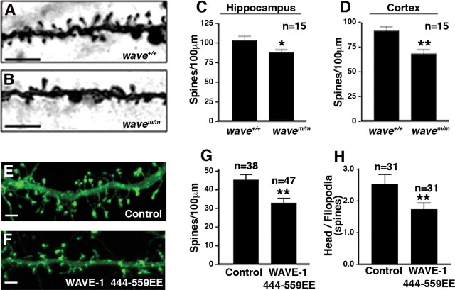
Figure 5.
Anchored WRP regulates spine density via WAVE-1 and Arp2/3. Hippocampal sections of wild-type (A) and mWAVE knock-in (B) mice after Golgi impregnation show dendritic segments of neurons. Quantitation of spine density (spines/100 μm) from hippocampus (C; area CA1) (*p = 0.0375 using two-tailed unpaired t test) and cortex (D; layer 1) (**p = 0.0015 using two-tailed unpaired t test). E, F, Fluorescence image of a section of dendrite showing the spine density in cultured hippocampal neurons expressing either YFP–actin (E) or WAVE-1 444–559EE and YFP–actin (F). G, Quantification of spine density (spines/100 μm) (**p = 0.0027 using two-tailed unpaired t test). H, Quantification of spine morphology (ratio of spines with heads/filopodial spines) (**p = 0.033 using two-tailed unpaired t test). All data are presented as mean ± SEM. Scale bars, 5 μm.

