
Figure 2. SREBP-2 activates miR-96/182/183 promoter.
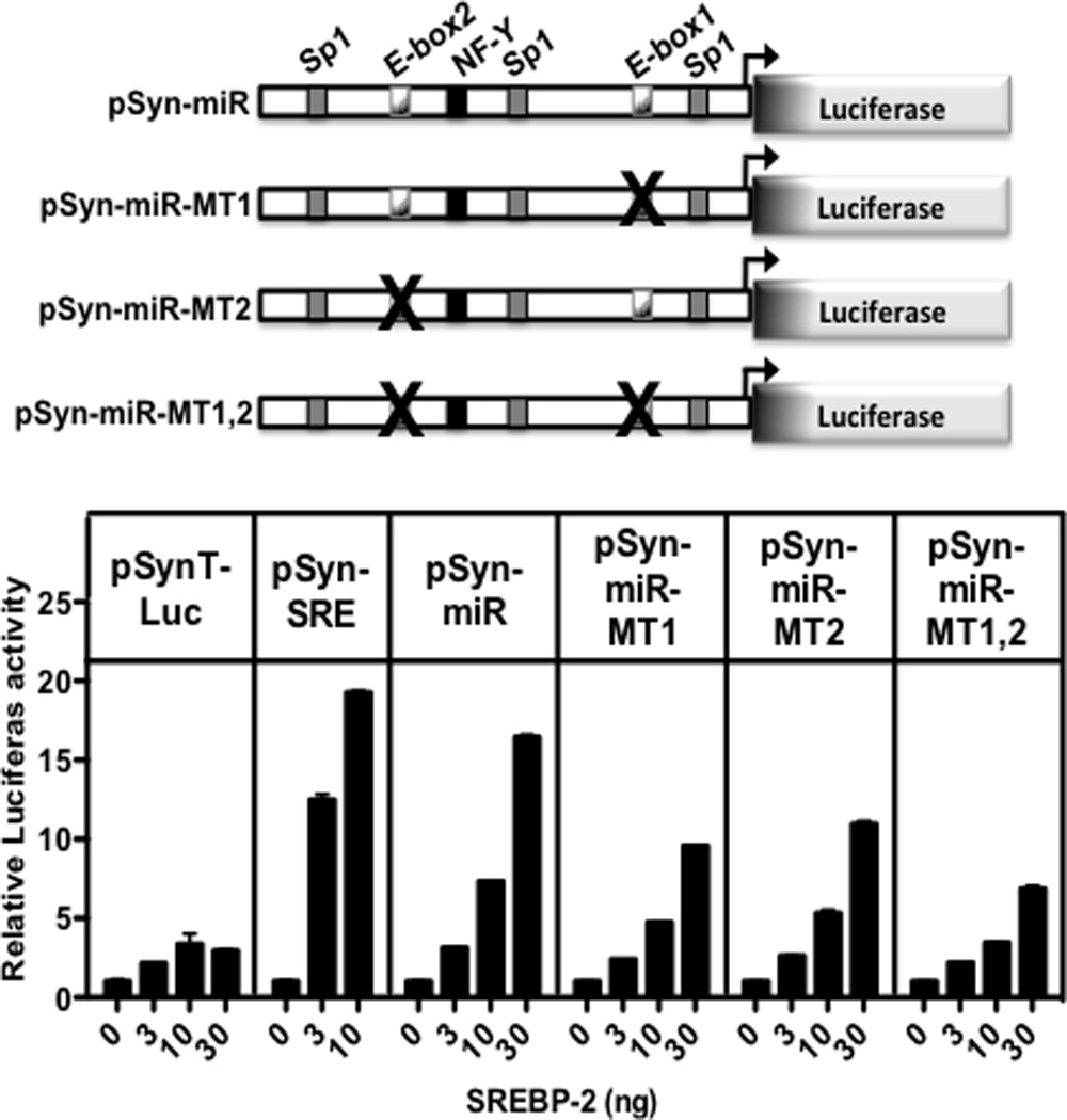
The putative promoter region shown in gray at the bottom of Fig. 1 was cloned upstream of luciferase in the control luciferase reporter as shown and described in Materials and Methods. Key putative transcription factor binding sites that are conserved between mouse and human (Fig. S3) are noted on the diagram of the sequence. (Top) There are two E-box motifs that are putative SREBP response elements and point mutations were engineered into each separately or in combination as noted by the “X”. (Bottom) Wild-type and the indicated mutant promoters were transfected into HEK-293T cells along with increasing amounts of an SREBP-2 expression vector as described in Materials and Methods. The negative and positive control promoters analyzed in parallel are shown as pSynTLuc and pSynSRELuc and are described elsewhere (Dooley et al., 1998). Luciferase activities were normalized to β-galactosidase that was expressed from an internal control co-transfected CMV- β-gal plasmid. Data are represented as mean +/− SEM. See also Figure S3.