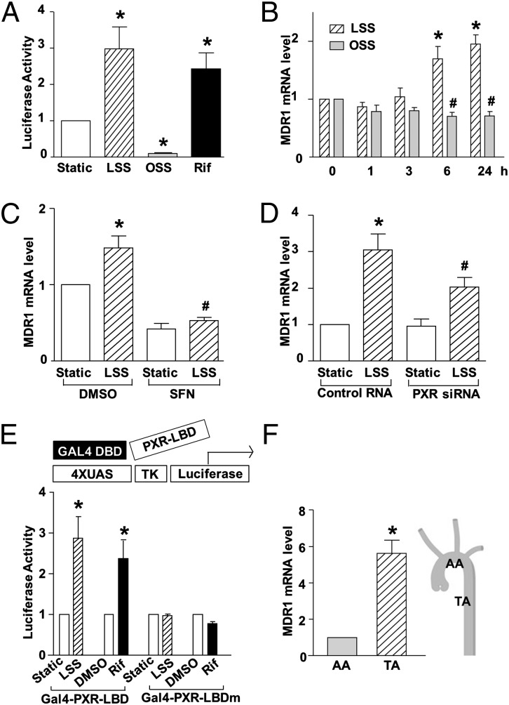
Fig. 1.
Shear stress regulates PXR activity in ECs. (A) BAECs were cotransfected with the PXR overexpression plasmid together with pCYP3A4XREM-362/+53, which is the plasmid for PXRE-driven luciferase reporter. Transfected cells were exposed to LSS or OSS or kept static for 18 h. Rifampicin (Rif, 20 μM) was used as a positive control. (B) MDR1 expression was analyzed by qRT-PCR in HUVECs exposed to LSS or OSS for 0, 1, 3, 6, and 24 h. (C and D) HUVECs were pretreated with SFN or DMSO for 24 h (C) or transfected with PXR siRNA or control siRNA for 48 h (D) and then exposed to laminar flow for another 6 h (C) or 24 h (D), mRNA levels of MDR1 were analyzed by qRT-PCR. (E) BAECs were transfected with the GAL4 reporter plasmid together with Gal4-PXR-LBD or Gal4-PXR-LBDmutant. Cells were then exposed to LSS or kept under static condition for 24 h. Rifampicin was used as a positive control. The results are expressed as fold change in luciferase activities compared with static control. Luciferase activity was assayed, normalized, and expressed as fold induction compared with static control. (F) MDR1 mRNA levels from intima at TA and AA were analyzed by qRT-PCR. Data are shown as mean ± SEM of three independent experiments. *P < 0.05 vs. control. #P < 0.05 vs. static (B) or control and with LSS treatment (C and D).

