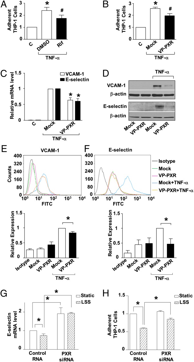
Fig. 3.
PXR activation ameliorates proinflammatory response in ECs. (A) HUVECs were pretreated by rifampicin (20 μM) or DMSO for 24 h before exposure to TNF-α (10 ng/mL, 4 h). Then ECs were incubated with fluorescence-labeled THP-1 cells (5 × 10⁵ cells per mL) for 30 min. THP-1 adhesions were counted under a fluorescence microscope. (B) Cells were infected with Ad-VP-PXR or mock infection before TNF-α stimulation. (C) VCAM-1 and E-selectin mRNA level was analyzed by using qRT-PCR in VP-PXR-infected or mock-infected ECs. (D) Protein levels of VCAM-1 and E-selectin were detected using Western blotting. (E and F) Surface expressions of VCAM-1 and E-selectin were examined using flow cytometry, and relative levels were quantified and expressed as bar graphs (Lower). (G) HUVECs were transfected with PXR or control siRNA and exposed 48 h later to LSS or static condition for 24 h. After stimulation with TNF-α for 2 h, RNA was extracted and analyzed by qRT-PCR. (H) Cells were stimulated with TNF-α for 4 h and incubated with THP-1 cells. THP-1 adhesions were counted; representative images are shown. Data are shown as mean ± SEM of three independent experiments. *P < 0.05 vs. control. #P < 0.05, Rif vs. DMSO (A) or VP-PXR vs. mock (B).

