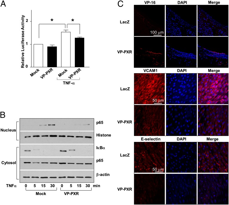
Fig. 4.
PXR inhibits NF-κB and vascular inflammation. (A) BAECs were cotransfected with a reporter plasmid NF-κB×5-luc together with a VP-PXR expression plasmid or vector control (pcDNA3.1). Data are shown as mean ± SEM of three independent experiments. *P < 0.05 vs. control. (B) Protein levels of p65 and IκBα in the nucleus and cytosol were analyzed using Western blotting in VP-PXR or mock-infected HUVECs after exposed to TNF-α. Histone and β-actin were used as internal controls for nuclear and cytosolic protein, respectively. (C) Rat carotid arteries were infected with Ad-VP-PXR or Ad-LacZ. Immunofluorescence staining shows overexpression of VP-PXR in endothelium 24 h after the infection. Expression of VCAM-1 and E-selectin after LPS challenge for 24 h was detected with en face staining. Nuclei were counterstained with DAPI. Microphotographs are representative of 3 rats in each group.

