Fig. 4.
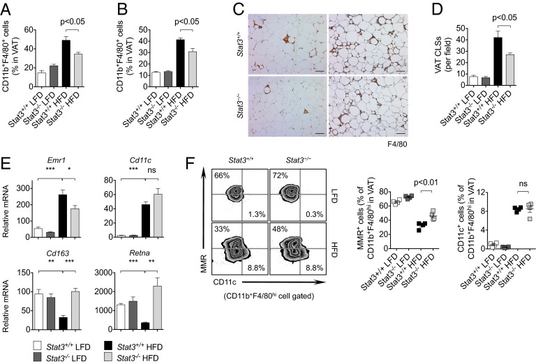
Stat3 in T cells regulates ATMs in DIO mice. (A) VAT-associated macrophages measured by flow cytometry in Stat3+/+ and Stat3−/− female mice on a HFD. n = 4 per group. (B) VAT-associated macrophages measured by flow cytometry in male mice on a HFD. n = 4 per group. (C) Histological evaluation of F4/80+ macrophages in VAT of female (Left) and male (Right) mice on a HFD. (Scale bars: 100 μm.) (D) Analysis of CLSs of F4/80+ macrophages in VAT of male mice on a LFD or HFD. n = 4 per group. (E) qRT-PCR analysis of VAT in mice on a LFD or HFD. n = 4 per group. (F) Flow cytometry analysis of M1 (CD11c) and M2 (MMR) macrophage markers in ATMs in VAT of mice on a LFD or HFD. Data are representative of two independent studies.

