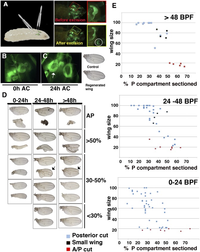
Fig. 1.
Method for surgical ablation in situ. (A) Representation of the method used to surgically remove sections of the wing discs in situ. (Right) An en-Gal4 UAS-GFP wing disc through the cuticle of the larvae before and after the excision. (B) An amputated Gal4-zfh2 LP30 UAS-GFP disc after the excision (48 h BPF). (C) The discs shown in B but 24 h later (arrow indicates regenerating disc). (Right) The adult wing differentiated from these discs. (D) Different examples of adult regenerated wings and control contralateral wings (upper wings). The discs were amputated at different times during the development. Depending on the size of the fragment amputated [shown as the percentage of the P compartment eliminated (Right)] and time past BPF we observed a range of regenerated adult wings phenotype. (E) Graphs comparing the adult wing sizes (y axis) with the percent of the P compartment eliminated (x axis) at different times during disc development. The red squares indicate discs in which we removed part of both A and P compartments. The black squares represent normally patterned small wings, arrows in D.

