Fig. 2.
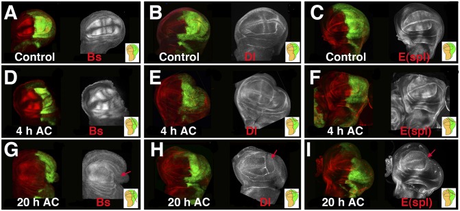
Regeneration induces changes in cell-fate commitment and patterns. Third instar wing imaginal discs stained with anti-Bs (A, D, and G), anti-Delta (B, E, and H), and anti-CD2 (C, F, and I) to visualize the expression of the E(spl)mβ-CD2 reporter in the control (A–C) and regenerating discs (D–I). (D, E, and F) At 4 h AC, the expression of Bs, Dl, and the E(spl)mβ reporter were similar to the controls compared with A–C. However, at 20 h AC, the expression of Bs (G), Dl (H), and E(spl)mβ-CD2 (I) defining the vein/intervein pattern was no longer evident. Near the wound edge, we observed a down-regulation of these markers (red arrows). Interestingly, the vein/intervein pattern also disappeared in the anterior compartment, although the cuts were only in the P compartment. Here and in the rest of the figures, Insets indicate the cutting lines and the region eliminated in each disc. Here, as in all of the disc figures, posterior is to the right, and it is marked in green by the expression of UAS-GFP driven by en-Gal4.

