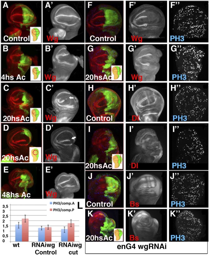
Fig. 3.
Evolution of Wg expression during wing disc regeneration. (A–G′) Expression of Wg, shown by anti-Wg (red in A–G) in en-Gal4; UAS-GFP third instar control discs (A–A′), regenerating discs at different times AC (B–E′) and in the en-Gal4 UAS-GFP /UAS-wg RNAi control (F–F′) and regenerating discs (G–G′). (H–K″) The en-Gal4 UAS-GFP /UAS-wg RNAi wing discs stained for anti-Dl (red in H and I) and anti-Bs (red in J and K). Mitotic cells were marked with Phospho-Histone H3 (blue in F–K). (B–B′) The expression of Wg was not altered at 0–4 h AC. (C–D′) At 20 h AC the expression of Wg disappears at the D/V boundary near the wound edge as well as in a section of the anterior wing margin (white arrows in C′ and D′). At this time the expression of the internal ring of Wg was partially (C–C′) or totally (D –D′) restored. (E–E′) At 48 h AC the expression of Wg was restored. (F–G′) Wing discs of en-Gal4 UAS-wg RNAi larvae displayed a strong down-regulation of Wg in the P compartment. (G–K″) Cell proliferation increase in the posterior compartment of regenerating discs with undetectable levels of Wg; see also L. The vein/intervein pattern defined by Dl (H–H′) and Bs (J–J′) in the control discs was disrupted in the en-Gal4 UAS-wg RNAi regenerating discs 20 h AC (I–I′ and K–K′). (L) Bar chart showing the average mitotic index in the posterior and anterior compartments of en-Gal4 UAS-GFP (wt control); en-Gal4 UAS-wg RNAi larvae raised at 17 °C and shifted to 25 °C for 40 h before being dissected (RNAiWg control); and regenerating discs of en-Gal4 UAS-wg RNAi larvae treated as in F–G (RNAiWg cut).

