Fig. 5.
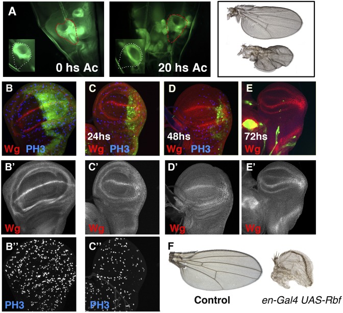
Evolution of disc regeneration in situ. (A) Image of a Gal4-zfh2 LP30 UAS-GFP amputated disc through the cuticle of the larvae after the excision, compared with the control discs (Inset). The disc was sectioned 48 h BPF. (Right) An image of the same disc but 20 h later. Note the smaller size of the amputated disc compared with the contralateral disc (35% smaller). The amputated disc is outlined in red, and the control disc is outlined in white. The adult wing differentiated from these discs are shown on the Right. Control (B–B″) and en-Gal4 UAS-Rbf280 UAS-GFP /Tub-Gal80ts (C–C″) third instar wing discs, stained for Wg (red in B–E) and PH3 (blue in B–D). Larvae were raised at 25 °C until 96 h AEL and shifted to 29 °C to block cell proliferation. Note the strong reduction of the number of mitotic cells in the P compartment, as assayed with PH3. (C–E) The en-Gal4 UAS-Rbf280 UAS-GFP /Tub-Gal80ts wing discs after 24 h (C), 48 h (D), and 72 h (E) at 29 °C. (F) Control wing and adult wing differentiated from the en-Gal4 UAS-Rbf280 UAS-GFP /Tub-Gal80ts larvae that were shifted to 29 °C at 96 h AEL and maintained at that temperature until the end of development.

