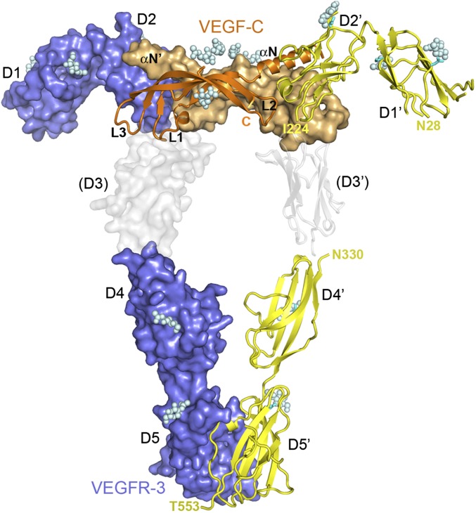
Fig. 1.
Crystal structures of the VEGF-C/VEGFR-3 D1-2 complex and the homodimer of VEGFR-3 D4-5. Shown are surface and cartoon representations with the two chains of VEGFR-3 in slate blue or yellow and the two chains of VEGF-C in orange or light orange. The VEGFR-3 complex, the D4-5 homodimer and our previous VEGF-C/VEGFR-2 D2-3 complex (PDB code 2X1X) were superimposed to the KIT/SCF complex (PDB code 2E9W). VEGFR-2 D3 is in gray. VEGFR Ig domains 1–5 (D1–D5), VEGF-C loops 1–3 (L1–L3), and the N-terminal helix (αN) are labeled. Glycan moieties are shown as spheres.

