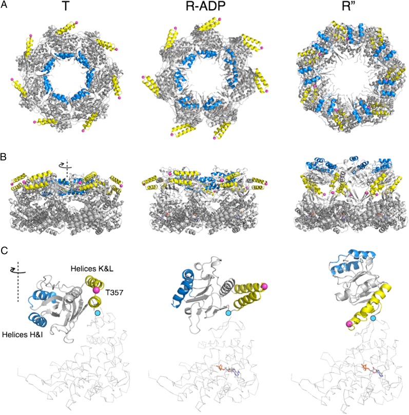
Fig. 7.
Conformational changes in apical domains between the T (PDB ID code 1XCK), R-ADP, and R″ (PDB ID code 1AON) states. (A) Top and (B) sides views of a single GroEL ring in (Left) T, (Center) R-ADP, and (Right) R″ states. Positions of helices H and I (blue), helices K and L (yellow), and T357 (Cαs as pink spheres) are highlighted to trace apical domain rotations. (C) Detailed views of one subunit from each state. The conformation differences between the intermediate domains (in cartoon) are the result of domain rotation around hinge 2 (cyan dots). ADP is shown in sticks. Note in particular that the major motion of helices K and L, a flip that brings T357 from its position on the external surface of GroEL to one on the internal surface, occurs during the R to R″ transition.

