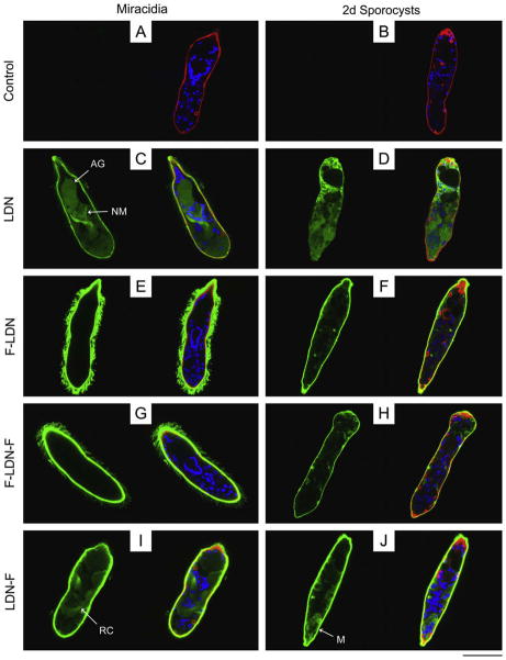
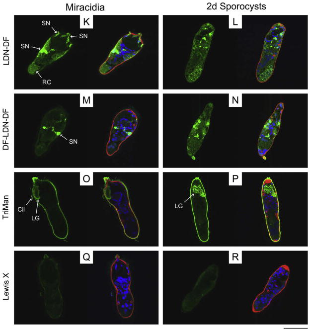
Fig. 1.
Localisation of glycotope expression in miracidia and primary sporocysts of Schistosoma mansoni. Confocal laser scanning microscopy was used to assess the localisation of schistosome-associated carbohydrate terminal structures in miracidia and 2-day (2d) in vitro-cultivated primary sporocysts. Fixed and permeabilised larvae were immunostained with monoclonal antibodies recognising GalNAcβ1-4GlcNAc (LDN; (C and D)), Fucα1-3GalNAcβ1-4GlcNAc (F-LDN; (E and F)), Fucα1-3GalNAcβ1- 4(Fucα1-3)GlcNAc (F-LDN-F; (G and H)), GalNAcβ1-4(Fucα1-3)GlcNAc (LDN-F; (I and J)), GalNAcβ1-4(Fucα1-2Fucα1-3)GlcNAc (LDN-DF; (K and L)), Fucα1-2Fucα1- 3GalNAcβ1-4(Fucα1-2Fucα1-3)GlcNAc (DF-LDN-DF; (M and N)), Manα1-3(Manα1-6)Manβ1-4GlcNAcβ1-4GlcNAcβ1-Asn (TriMan; (O and P)) and Galβ1-4(Fucα1-3)GlcNAc (Lewis X; (Q and R)). Control larvae were exposed to secondary antibody without previous primary antibody treatment (A and B). Panels include paired micrographs depicting glycotope expression (green) alone and merged with counterstained actin (e.g., muscles, flame cells; red) and DNA (e.g., nuclei; blue). Approximate scale is represented in the lower right corner (bar = 50 μm). AG, apical gland; Cil, cilia; LG, lateral gland; M, sporocyst matrix; NM, neural mass; RC, interepidermal ridge cyton; SN, sensory nerve.


