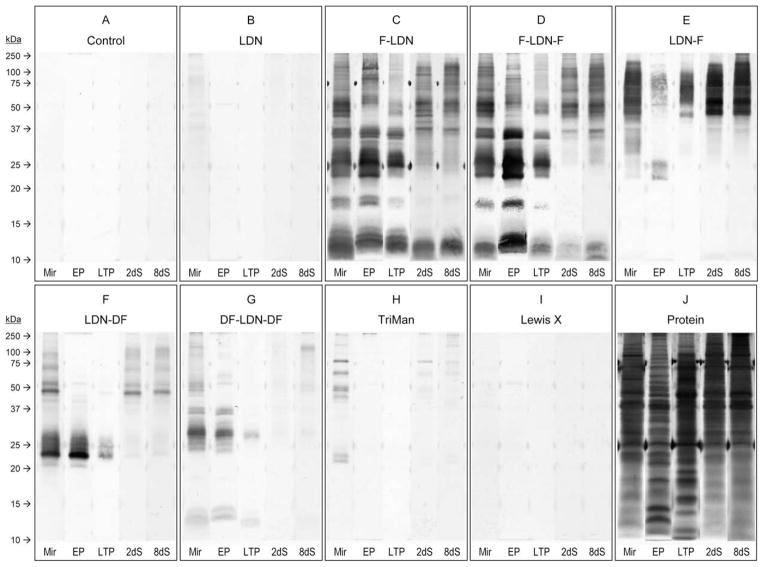
Fig. 4.
Western blots revealing glycotope expression amongst whole-body larval extracts, epidermal plate glycoproteins and parasite culture supernatants containing larval transformation proteins. Whole-body extracts of miracidia (Mir) and primary sporocysts cultivated 2 and 8 days (2dS and 8dS, respectively), as well as ciliated epidermal plate extracts (EP) and parasite culture supernatants containing larval transformation proteins (LTP), were SDS–PAGE-fractionated and immunoblotted using anti-glycotope monoclonal antibodies that recognise GalNAcβ1-4GlcNAc (LDN; (B)), Fucα1-3GalNAcβ1-4GlcNAc (F-LDN; (C)), Fucα1-3GalNAcβ1-4(Fucα1-3)GlcNAc (F-LDN-F; (D)), GalNAcβ1-4(Fucα1-3)GlcNAc (LDN-F; (E)), GalNAcβ1-4(Fucα1-2Fucα1-3)GlcNAc (LDN-DF; (F)), Fucα1-2Fucα1-3GalNAcβ1-4(Fucα1-2Fucα1-3)GlcNAc (DF-LDN-DF; (G)), Manα1-3(Manα1-6)Manβ1-4GlcNAcβ1-4GlcNAcβ1-Asn (TriMan; (H)) and Galβ1-4(Fucα1-3)GlcNAc (Lewis X; (I)). Control blots were exposed to the secondary antibody without previous primary antibody incubation (A), and total protein was visualised in-gel by silver stain (J).

