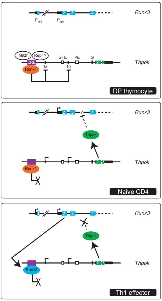
Figure 3. Transcriptional plasticity at the Thpok locus.

The Runx3 and Thpok loci are schematically represented in three separate boxes. For each gene, transcription start sites are shown as arrows and exons as numbered cyan or green boxes; non-coding exonic sequences are depicted as thinner black rectangles. Note the two alternative promoters and first exons (1a and 1b) of Thpok. Boxes (top) indicate the distal regulatory element (DRE, which includes or overlaps with the silencer [27,31]) the general T lymphoid element (GTE), the proximal enhancer (PE), and a conserved sequence (G) that includes a Gata3 binding site [13]. The distal (Pdis) and proximal (Pprox) Runx3 promoters are indicated upstream of exons 1 and 2, respectively. In DP thymocytes (top), Thpok is not transcribed because of reversible repression involving Runx1, Mazr and potentially unknown other repressors (Repr ?). The Runx3 distal promoter, that gives rise to protein-producing transcripts, is not active. In CD4 SP thymocytes and naïve CD4 T cells (middle), Thpok is expressed and represses Runx3 through so far unknown mechanisms (dotted line); note that this does not prevent transcription from the proximal promoter [23]. Although Runx1 is expressed and bound to the silencer, it does not repress Thpok, possibly because of the absence of the other repressors (see text for discussion). In Th1 effectors (bottom), Thpok is expressed but no longer represses the Runx3 distal promoter. Conversely, Runx3 fails to repress Thpok; it is shown as bound to the silencer by analogy with Runx1 in resting cells (see text for discussion). Drawings are not on scale.
