
Figure 1. Osteochondral defect.
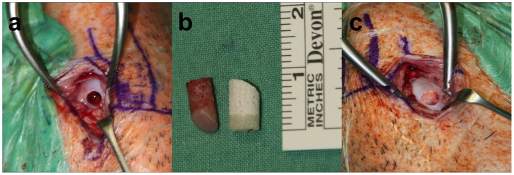
In all animals, a 6×10 mm cylindrical osteochondral defect in the medial femoral condyles of both knee joints was surgically created with a cylindrical chisel (a). The osteochondral graft (left) from the defect was measured and then the biphasic scaffold (right) was cut to the respective length of the graft prior to implantation (b). The cut scaffold was then supplemented with the respective supplement and implanted into the condyle (c).