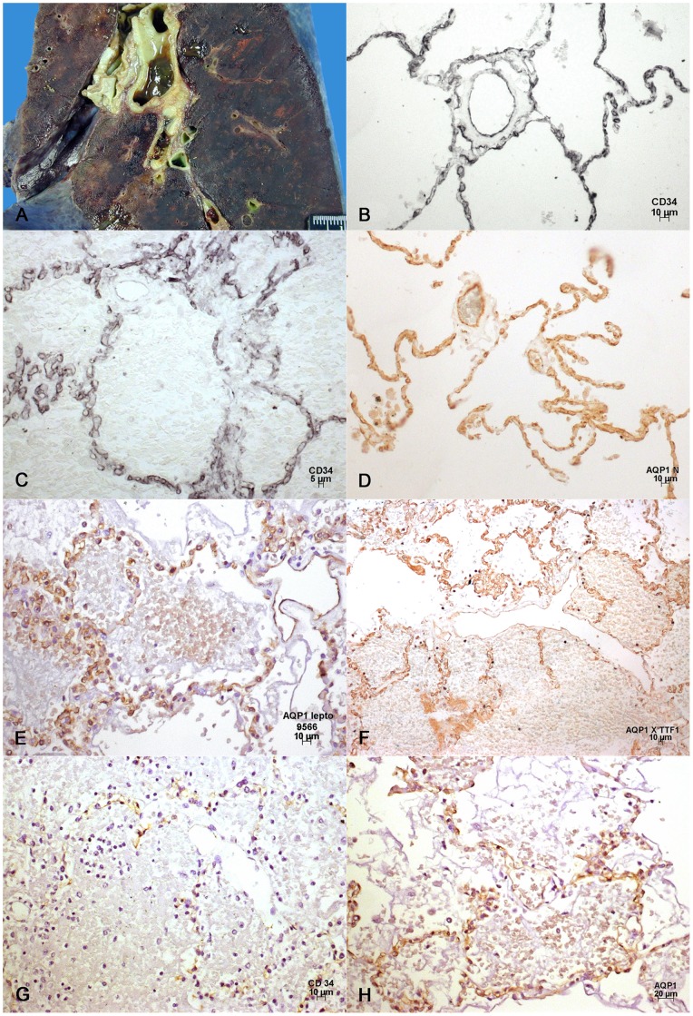
Figure 2. Gross feature and immunohistochemical analysis of microcirculation of leptospirotic lungs: A: Macroscopic aspect of the hemorrhagic pneumopathy in leptospirosis.
Confluent hemorrhagic areas are present in the lung parenchyma. B: Microcirculation of the normal human lung. The capillary network is delineated in black, as well as the endothelium of a small branch of the pulmonary artery. IHC CD 34, DAB-Nickel. C: Human lung in leptospirosis. The capillary vessels are frequently dilated, with small gaps and areas of reduced and/or absent expression of CD 34. IHC CD 34, DAB-Nickel. D: Aquaporin 1 delineates the walls of the microcirculatory vessels in the normal human lung. It is also expressed in the endothelium of a small branch of the pulmonary artery. IHC, DAB. E: Aquaporin 1 expression is mostly preserved in areas of edema and apparent red blood cell deposits in human lung in leptospirosis. IHC Aquaporin 1, DAB. F: Capillaries of the pulmonary microcirculation express aquaporin 1 both at the more preserved periphery and inside the area of intraalveolar edema and apparent red blood cells extravasate. IHC Aquaporin 1. G and H: Both images were taken from similar regions of the slide. G shows CD34 reduced expression in areas of edema and hemorrhage and H the relative preservation of capillary expression of aquaporin 1. IHC, DAB.

