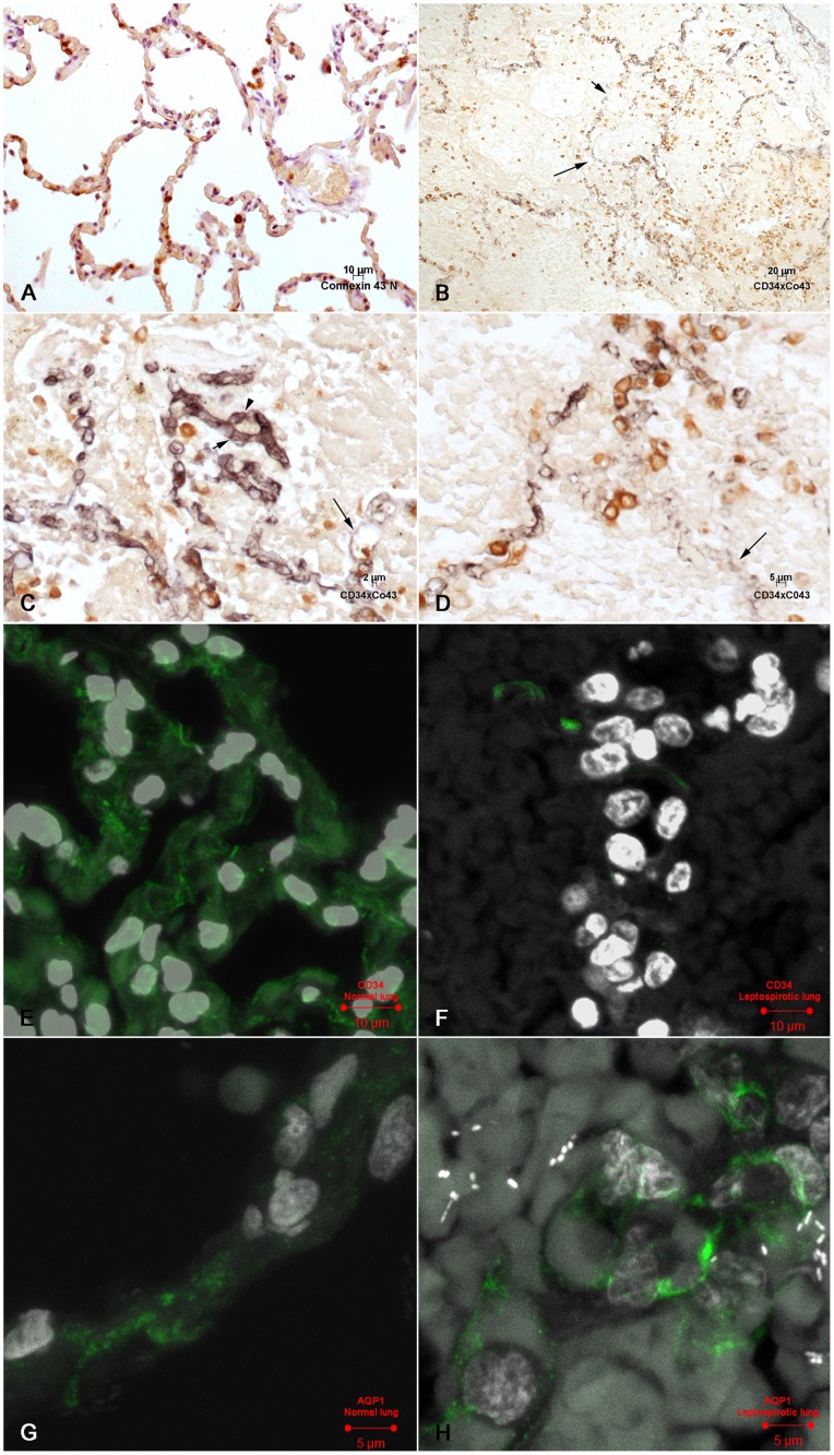
Figure 3. Immunohistochemical and confocal analysis of microcirculation of leptospirotic lungs: A: Connexin 43 is expressed in cells of the alveolar epithelial lining of a normal human lung.
IHC, DAB. B: Lung in human leptospirosis. Connexin 43 is expressed both in pneumocytes of the epithelial lining and in those inside area of alveolar edema. Capillary with partial expression of CD34 in the endothelial membrane(long arrow) and a gap are present in the alveolar epithelium (short arrow). IHC, double labelling. C: Lung in human leptospirosis: Connexin 43 is expressed in the cytoplasm of isolated pneumocytes of the alveolar epithelial lining. CD34 is expressed in the capillary membranes. The short arrow and arrow head show apparently preserved capillary junctions. The long arrow points to a dilated capillary with reduced CD34 expression. Alveolar lumen is filled with plasma material and shadows of structures that might be interpreted as red blood cells. IHC, double labelling. D: Lung in human leptospirosis: Preserved expression of connexin 43 in groups and/or isolated pneumocytes. CD 34 shows an area with an almost complete lack of expression in the capillary membrane (long arrow) and irregular expression in the other capillaries. IHC, double labelling. E: Confocal microscopy of normal lung. The capillary walls are delineated by immunofluorescence. CD34 expression. F: Confocal microscopy in the human lung in leptospirosis: A few segments of capillary walls are delineated by immunofluorescence. CD34 expression. G: Confocal microscopy of normal human lung. Aquaporin 1 appears as fluorescent granules, probably in the cytoplasm of endothelial cells. H: Human lung in leptospirosis. Endothelial fluorescent granules are still present, probably in the cytoplasm of endothelial cells.

