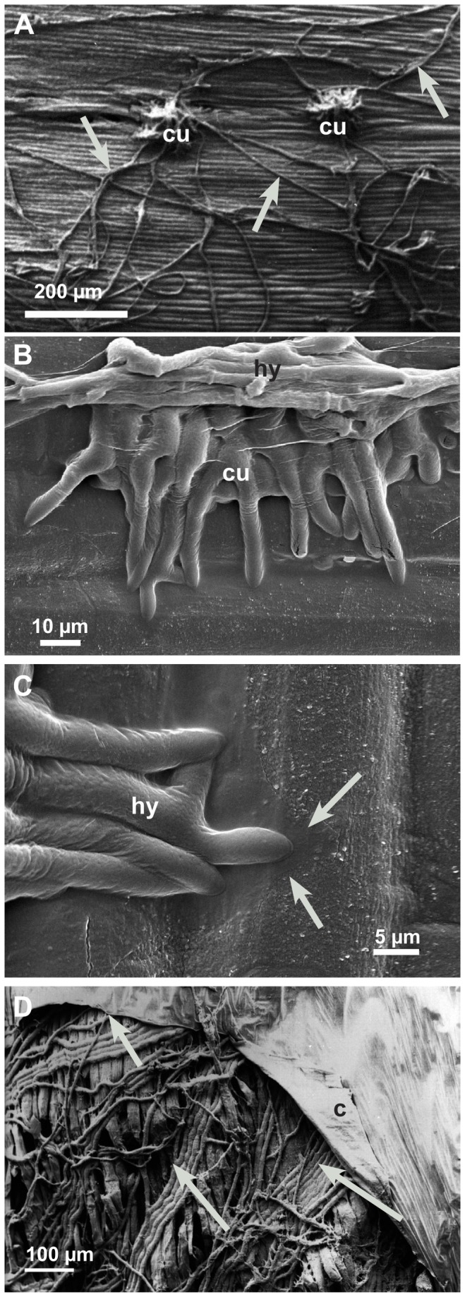
Figure 2. Development of early infection structures (12-24 hpi).

Infection cushions, running hyphae on the epidermis of hypocotyls, and subcuticular hyphae investigated by scanning electron microscopy. A: Dome-shaped infection cushion (cu) and running hyphae (arrows); conventional SEM. B: Young infection cushion (cu) overgrown by running hyphae (hy); LTSEM. C: Detail of Figure 2B. Hyphae (hy) of the infection cushion attached to the cuticle of the epidermis. Hyphal exudates (arrows) covering wax crystals of the cuticle in LTSEM. D: After penetration, when the cuticle (c) is detached from the epidermal layer the parallel growing subcuticular hyphae (arrows) become visible in conventional SEM.
