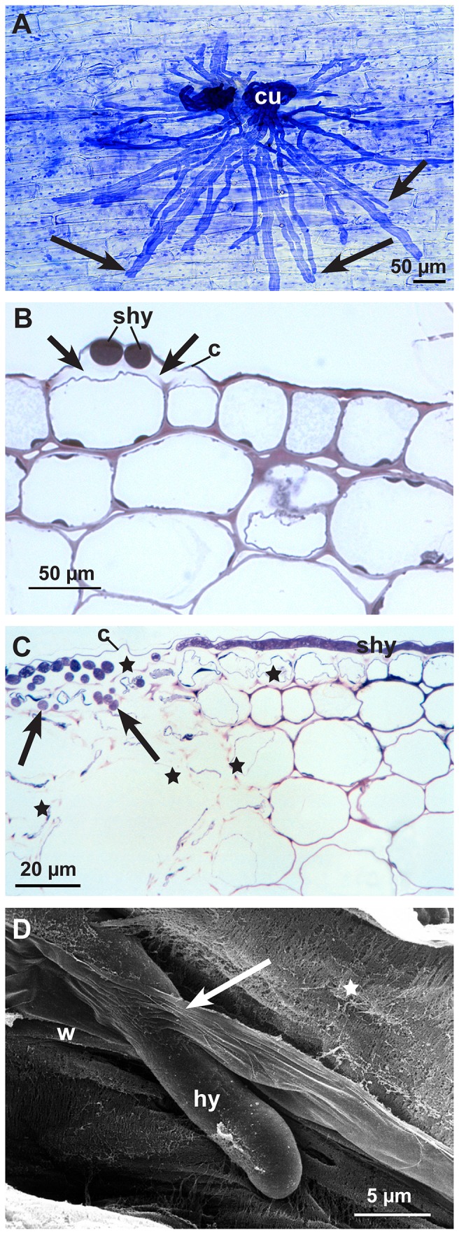
Figure 3. Infection process of the early (12-24 hpi) and advanced infection stages (36-48 hpi).

Subcuticular hyphae and hyphal growth starting in epidermal and adjacent cortical parenchyma cells. A: Subcuticular hyphae (arrows) spreading fan-like from two infection cushions (cu). Light micrograph of an epidermal strip stained with Coomassie blue 12-24 hpi. B: Cross section of subcuticular hyphae (shy) near hyphal tips in the abaxial cell wall. Under the cuticle (c) around hyphae the loss of contrast and the widening of the cell wall indicating cell wall degradation (arrows). Light micrograph stained with toluidine blue 12-24 hpi. C: Cross section of infected hypocotyl with subcuticular hyphae (shy) under the cuticle (c) and hyphae growing deeper into the host tissue (arrows). Cell walls and cytoplasm of the epidermal cells are completely destroyed as well as parts of the cortical parenchyma (stars). Light micrograph stained with toluidine blue 36-48 hpi. D: Scanning electron micrograph, 36-48 hpi. In a cortical parenchyma cell the fibrillar (stars) and lamellar (arrow) structure of the degrading cell wall (w) becomes visible around an invading hyphae (hy) by conventional SEM.
