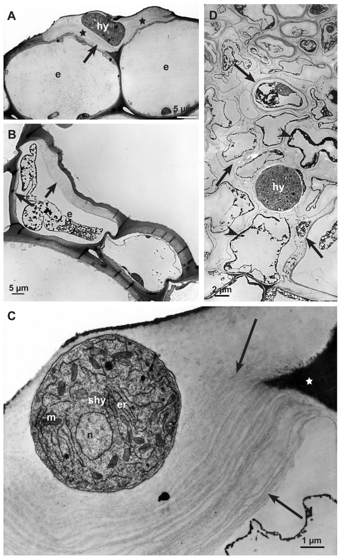
Figure 4. Degradation process of the cell wall matrix in early (12-24 hpi) and advanced (36-48 hpi) infection stages.
Transmission electron micrographs of cross sections of sunflower hypocotyls: A: The abaxial epidermal cell wall around a subcuticular hypha (shy) 12-24 hpi showing degraded cell wall matrix (stars). Cytoplasm of the epidermal cells (e) still intact. B: Destruction process of cell wall in dead host cells starting from the inner side of the cell wall (arrows) of an epidermal cell (e) 12-24 hpi. C: Detail of a degraded abaxial epidermal cell wall around a subcuticular hypha (shy); non-degraded part of the plant cell wall (star), cell wall matrix degraded, residues of cellulose layers (arrows), nucleus (n), endoplasmic reticulum (er) and mitochondrium (m) of the fungal cell. D: During the advanced infection stage (36-48 hpi) a single hypha (hy) in a large area of the necrotic cortical parenchyma. Only residues of thin cell wall layers (arrows) and dark staining residues of cytoplasm are left (arrowheads).

