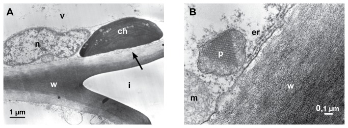
Figure 6. Reaction of the host cells.

Transmission electron micrographs (12-24 hpi). A: A chloroplast (ch) changing contrast, the matrix appear electron dense. The brightening of the inner cell wall layer is probably the beginning of the degradation of the cell wall (arrow), nucleus (n), vacuole (v), intercellular space (i) and cell wall (w). B: A cortical parenchyma cell with peroxisome (p) containing protein-crystal (probably catalase), mitochondrium (m), endoplasmic reticulum (er) and cell wall (w).
