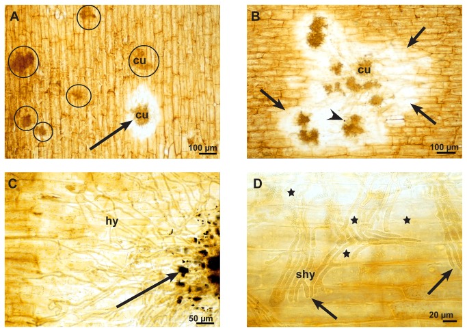
Figure 7. Secretion of oxalic acid in the infection process of S. sclerotiorum.
Light micrographs of the early, advanced, and late stages of infection after histochemical staining for calcium oxalate of fresh epidermal strips that resulted in a yellow-brownish colour of tissue and a brown-black colour of the compact oxalate crystals. A: At the early infections stage (12-24 hpi) the epidermal layer around one infections cushion (cu) is bleached (arrow), while most of the infection cushions (circles) do not show any influence on the epidermal layer. B: In the advanced stage (36-48 hpi) there is bleaching of the epidermal layer (arrows) around all infection cushions (cu) and only a single dark stained particle of oxalate was visible (arrowhead). C: At the late stage (72 hpi) there is not only bleaching, but calcium oxalate crystals accumulate around the infection cushions (arrow). D: Detail of a late stage at the infection front. The bleaching effect (stars) did not become visible at the hyphal tips (arrows), but along the older parts of the subcuticular hypha (shy).

