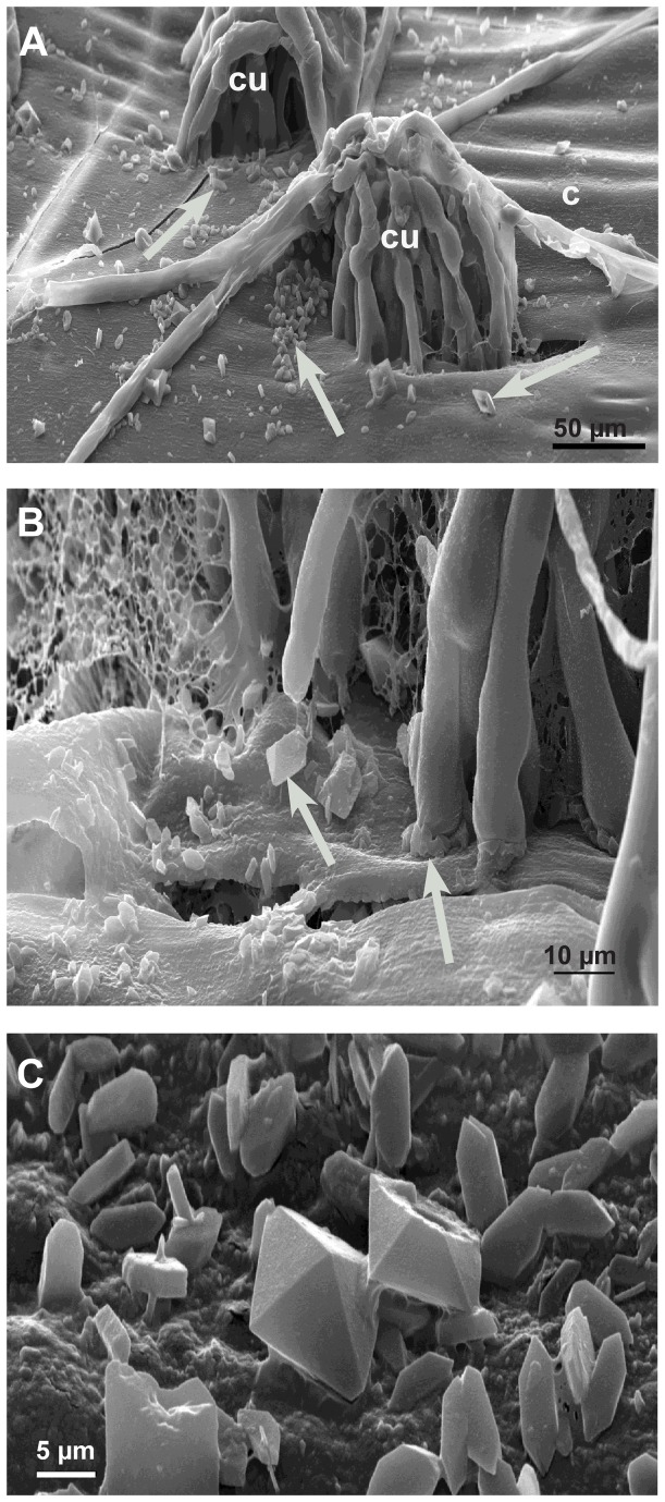
Figure 8. Calcium oxalate accumulation in the late infection stages (72 hpi) by LTSEM.
Scanning electron micrographs: A: On the detached cuticle (c) calcium oxalate crystals appear (arrows) around the infections cushions (cu) B: Detail of Figure 8A showing the accumulation of crystals (arrows). C: Detail showing the variation of calcium oxalate crystals.

