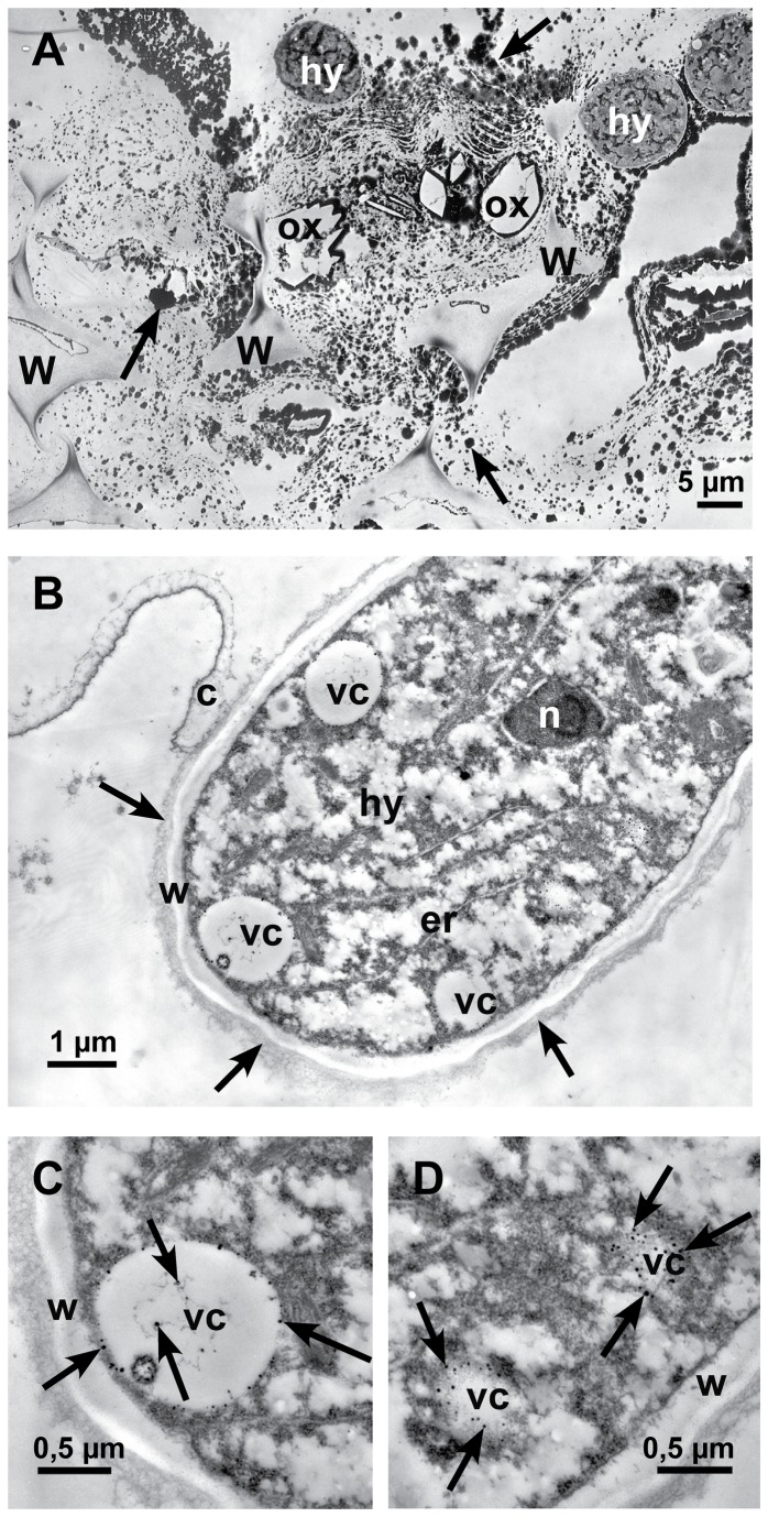
Figure 9. Localization of calcium by potassium pyroantimonate precipitation.
Transmission electron micrographs of cross sections of sunflower hypocotyls. A: In the late infection stage (72 hpi), near an infection cushion, dark staining precipitates (arrows) are detectable all over in the destroyed tissue, around hyphae (hy) and oxalate crystals (ox) (crystals itself were lost during sectioning). Only residues of cell walls (w) are left. B: In the early infection stage 12-24 hpi, the longitudinal section of a hyphal tip (hy) that just penetrated the cuticle (c) is showing vesicles (vc) with small dark stained calcium precipitates. Around the cell wall of the hypha an electron dense, fibrous matrix is visible (arrows), nucleus (n), endoplasmic reticulum (er), cell wall (w) and vesicles (vc) with calcium precipitates. C: Detail of Figure 9B: Cross section of a vesicle (vc) with dark stained calcium precipitates (arrows) and cell wall (w). D: Detail of Figure 9B: Tangential section of vesicles (vc) with dark precipitates of calcium (arrows) and cell wall (w).

