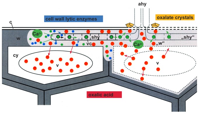
Figure 12. Infection model illustrating the complex interaction between S. sclerotiorum and its host cells sunflower.
Scheme of two epidermal cells infected by hyphae of S. sclerotiorum. After penetration of the cuticle (c) by appressorial hyphae (ahy), subcuticular hyphae (shy) grow in the outer epidermal wall layer (w) and live on the cell wall matrix (intact wall in dark grey, degraded cell wall matrix in light grey (“w”). Subcuticular hyphae (shy) secrete cell wall lytic enzymes (blue dots) and oxalic acid (red dots) that do not kill the host cells immediately (intact cell compartments in continuous lines; degraded cell compartments in broken lines). While the enzymes degrade the matrix of the host cell walls and set free calcium (green dots), oxalic acid (red dots) is metabolized by the host cells and stored in the host cell vacuoles (v). Calcium is taken up by the growing hyphae in vesicles (vc) and translocated back to the older part (“shy”) to prevent un-physiological high calcium concentrations. Finally, host cells die (broken lines), because the degradation of cell walls advances and the concentration of oxalic acid rise. Oxalic acid (red dots) and also vacuolar enzymes of the host cells are set free and an autolytic degradation start from the inner side of the host cell walls contributing to the development of necrotic tissue. After death of the senescent hyphae (broken lines), calcium is again set free and is reacting with oxalic acid building stable calcium oxalate crystals (yellow) in the necrotic tissue around the functionless appressorial hyphae (ahy) of the infection cushion.

