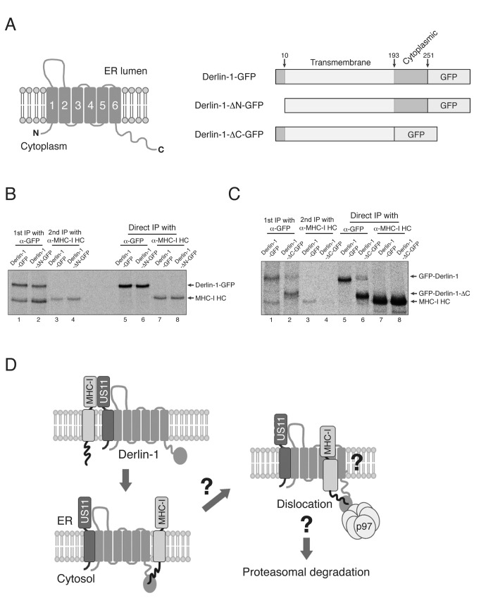
Figure 6. The C-terminal cytosolic region of Derlin-1 binds MHC-I molecules during US11-induced ERAD.
(A) Proposed membrane topology of Derlin-1 (left) and a schematic representation of the GFP-conjugated Derlin-1 constructs (B) The N-terminal cytosolic region of Derlin-1 is not required for binding to MHC-I molecules. U373MG-US11 cells were transfected with Derlin-1-GFP or Derlin-1-ΔN-GFP, metabolically labeled for 1 hr, lysed in 1% digitonin, and subjected to immunoprecipitation with anti-GFP antibody. The precipitate was then boiled in SDS/DTT-containing buffer, diluted 10-fold in 1% NP-40, and subjected to a second round of immunoprecipitation with mAb HC10, which reacts with the denatured form of MHC-I heavy chains. (C) Deletion of Derlin-1 C-terminal region abolishes its interaction with MHC-I molecules. U373MG-US11 cells were transfected with Derlin-1-GFP or Derlin-1-ΔC-GFP, metabolically labeled for 1 hr, lysed in 1% digitonin, and subjected to immunoprecipitation with anti-GFP antibody. The precipitate was then boiled in SDS/DTT-containing buffer, diluted 10-fold in 1% NP-40, and subjected to a second round of immunoprecipitation with mAb HC10. All experiments were performed multiple times with similar results, and the data shown are representative of all results. (D) Working model describing the cytosolic interaction between Derlin-1 and MHC-I molecules during US11-induced ERAD. See the DISCUSSION section for a detailed explanation.

