
Figure 1. TFAM Augments CpGA DNA-induced Splenocyte TNFα Release which Is Highly Dependent upon the Presence of Plasmacytoid Dendritic Cells.
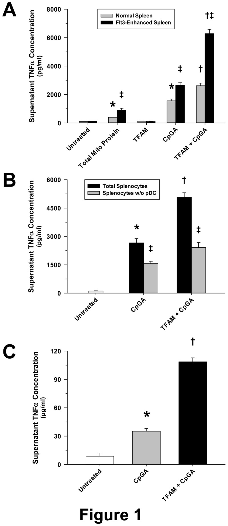
The presented data was derived from at least 5 independent experiments. (A) As previously observed for other cytokines in different cell preparations [2,3], native mitochondrial protein (5 µg/ml) induced significant TNFα release from cultured mouse Flt3L-expanded splenocytes (1 × 10⁶ cells/ml) 24 hours post-treatment. CpGA DNA (0.3 µM), which is akin to that naturally associated with mitochondrial protein, promoted splenocyte TNFα release which was significantly amplified by co-incubation with recombinant human TFAM (5 µg/ml) despite that having no effect when treated alone. Compared to splenocytes obtained from a normal spleen, TNFα release was markedly increased following all treatments when the constituent pDC population was expanded by Flt3L over-expression as employed in the present study. (B) Selective removal of pDCs from matching Flt3L-expanded splenocyte preparations notably reduced the TNFα release induced by CpGA DNA (0.3 µM) ± TFAM (5 µg/ml) 24 hours post-treatment. (C) pDCs (1 × 10⁶ cells/ml) isolated from similar mouse splenocyte preparations demonstrated comparably minimal yet significant TNFα release 24 hours after CpGA DNA (0.3 µM) ± TFAM (5 µg/ml) treatment (*p < 0.01, relative to no treatment; † p < 0.01, compared to CpGA DNA treatment alone, and ‡ p < 0.01, relative to matching normal splenocyte treatments).