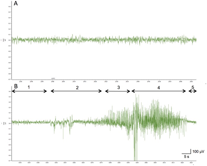
Figure 3. Ictal electroencephalogram recorded in CQ-injected NER.
A recording electrode was implanted into the hippocamal CA3 region of NER. Two days later, vehicle (A) or clioquinol (30 mg/kg) in vehicle (B) was i.p. injected into NER and then the behavior was observed in the home cage until seizures were observed. Note that no seizure was observed in vehicle-treated NER. The basal activity before seizures (1) and the stages of seizures were observed as follows: beginning of aberrant activity on electroencephalogram without behavioral changes (2); asymmetric wave with myoclonus (3); high-voltage wave with tonic-clonic convulsion (4); low-voltage wave with postictal flaccidness (5). The component of γ-wave (7–9 Hz) (2, 6.9%; 3, 19.4%; 4, 15.4%; 5, 10.0%) became rhythmic during seizures as reported previously [26].

