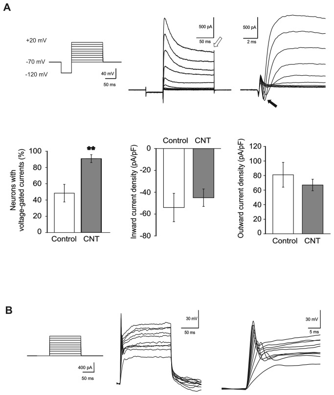
Figure 2. MWCNTs boost the functional maturation of spinal neurons.
(A) Top left, voltage-clamp stimulation protocol to test the presence of voltage-dependent currents. Top middle (and right, in extended time scale), typical recordings from a spinal neuron displaying voltage-dependent currents. Note the presence of both outward (open arrow, middle panel; K+) and inward (filled arrow, right panel; Na+) voltage-dependent currents. The fraction of neurons displaying voltage-dependent currents is considerably higher in CNT neurons with respect to controls (bottom left; **: P<0.01), while current density is similar for inward and outward currents in both culturing conditions (bottom middle and right). (B) Left, current-clamp stimulation protocol to test the neuronal ability to generate action potentials. Middle (and right, in extended time scale), example of action potentials generated by a spinal neuron.

