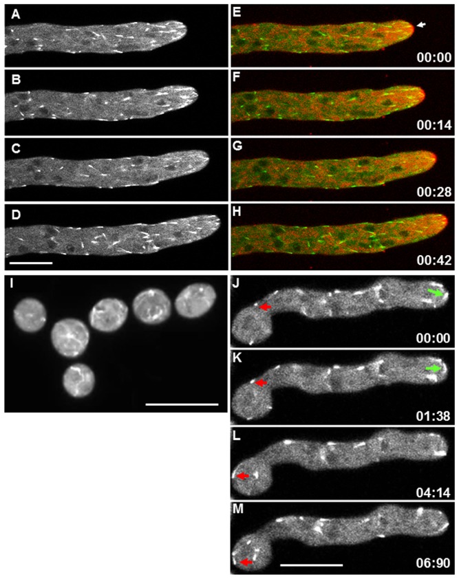
Figure 1. Distribution of MTB-3-GFP comets in different developmental stages of N. crassa.
(A–D) Time lapse images of MTB-3-GFP in mature hyphae. (E–H) merged images of MTB-3-GFP and membranes stained with 5 µM FM4-64 in mature hypha. The Spitzenkörper is indicated by a white arrow. (I) Conidia expressing MTB-3-GFP. (M–P) Time lapse images of MTB-3-GFP in germlings. Red arrows point to MTB-3-GFP comets that move in retrograde direction and green arrows point to comets that move in anterograde direction. Time scale is in min∶sec. Scale bars = 10 µm.

