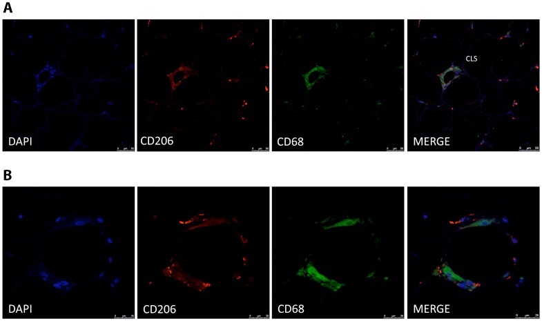
Figure 2. Immunofluorescence detection of CD206 and CD68 in human adipose tissue crown-like structures.
Co-staining of adipose tissue macrophages in “crown-like structures” demonstrates co-localization of CD206 (red) and CD68 (green) proteins in most of the cells (A). Close-up view of a CLS (B) The counterstaining of nuclei (DAPI) is shown in blue. Images are representative of adipose tissue preparations collected from three subjects. Scale bar, 50 µm (A) and 25 µm (B).

