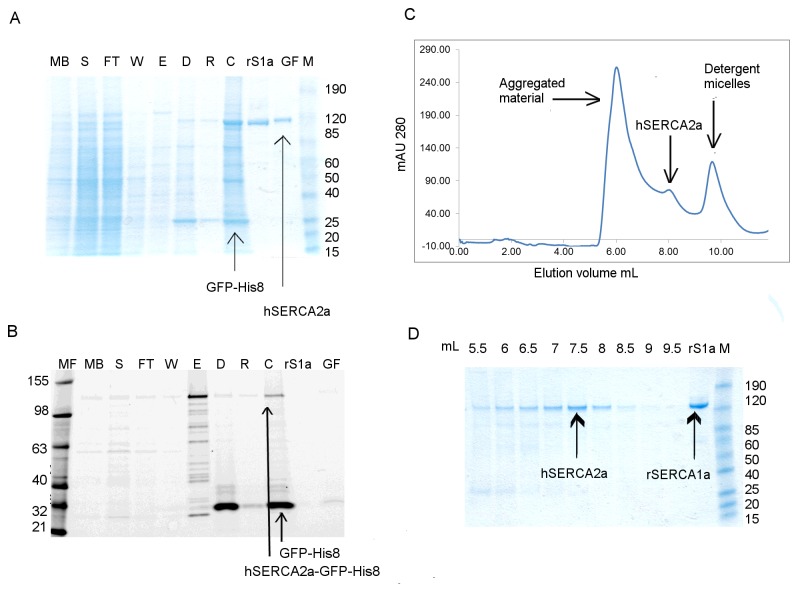
Figure 3. Purification of hSERCA2a-GFP-His8 using Ni-NTA affinity chromatography.
Purification was done in the presence of DDM only throughout all steps, including SEC. Protein was obtained using rich media. A. Coomassie stained SDS-PAGE gel; B. In gel fluorescence 12% Tris-Glycine SDS-PAGE gel. MF- fluorescent protein ladder; MB- diluted membrane fraction; S- solubilised fraction; FT- flow-through after binding; W- wash fraction; E- elution; D- sample after cleavage with TEV protease and dialysis; R- sample after Ni-NTA rebinding after tag cleavage; C- sample concentrated using 50 kDa cut-off filter concentrator, before gel filtration; S1a- rabbit SERCA1a; GF- fraction containing human SERCA2a after gel filtration; M- prestained protein ladder. C. HPLC-SEC profile for hSERCA2a purified using Ni-NTA super-flow resin. D. Coomassie stained 4-12% Tris-Glycine SDS PAGE gel for SEC fractions obtained for purification of hSERCA2a using Ni-NTA super-flow resin.

