
Figure 3. In vivo specific fluorescence imaging in the same mouse body.
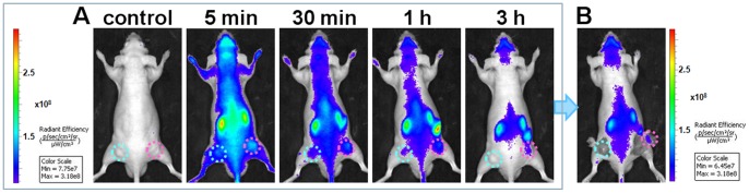
Images were acquired after an intravenous injection of Cy5-S6 in a nude mouse simultaneously bearing an A549 tumor (pink circles) and a Tca8113 tumor (cyan circles). (A) Time-lapse fluorescence imaging. (B) Imaging at 3 h postinjection with the tumor tissues uncovered.