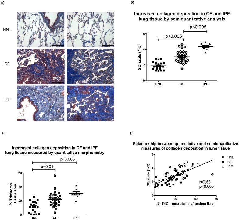
Figure 2. Increased peripheral lung fibrosis in cystic fibrosis (CF).
A) Histochemical staining for fibrosis (Masson trichrome stain, collagen shown in blue) in human normal lung (HNL), cystic fibrosis (CF) and idiopathic pulmonary fibrosis (IPF). Images are shown at approximately 100× from 2 separate HNL, CF and IPF specimens (bar = 250 µm). B) Comparison of collagen deposition (Masson trichrome stain) by semiquantitative analysis and C) quantitative morphometry. D) Correlation between quantitative morphometry (% collagen deposition) and semiquantitative analysis (1–5 scale).

