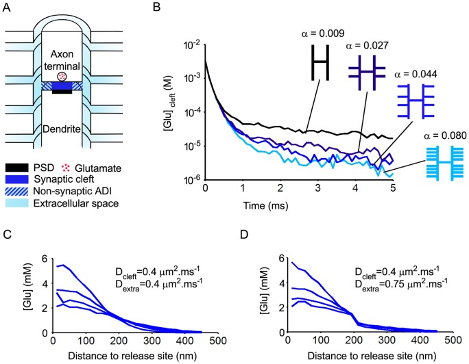
Figure 1. Effects of diffusion space geometry on the time course of glutamate in the synaptic cleft.
A. Three-dimensional representation (half view, not to scale) of the synapse model. The diffusion space includes the axon-dendrite interface (ADI, radius: 500 nm ; width: 12 nm) divided into a synaptic cleft bordered by the PSD (radius: 200 nm) and a peripheral non-synaptic part, the extracellular space bordering the axon and the dendrite (width: 20 nm) and four escape routes orthogonal to the synapse axis (width of escape routes: 20 nm). Glutamate release occurs at the center of the ADI. B. Effects of model porosity. The porosity α is gradually increased by successive additions of escape routes. Each trace is an average of 5 trials. Glutamate exit from the cleft is strongly accelerated by the incorporation of the first two proximal escape routes. Further additions of escape route have less prominent effects. C and D. Effects of extrasynaptic diffusion coefficient (Dextra) on spatio-temporal glutamate concentration profiles in the ADI. Traces were obtained at different time intervals after release (0.02 ms to 0.05 ms, each trace is the average of 5 trails). Increasing Dextra from 0.4 µm2.ms−1 to 0.75 µm2.ms−1 lowers glutamate concentrations in the non-synaptic part of the ADI but does not affect cleft glutamate content.

