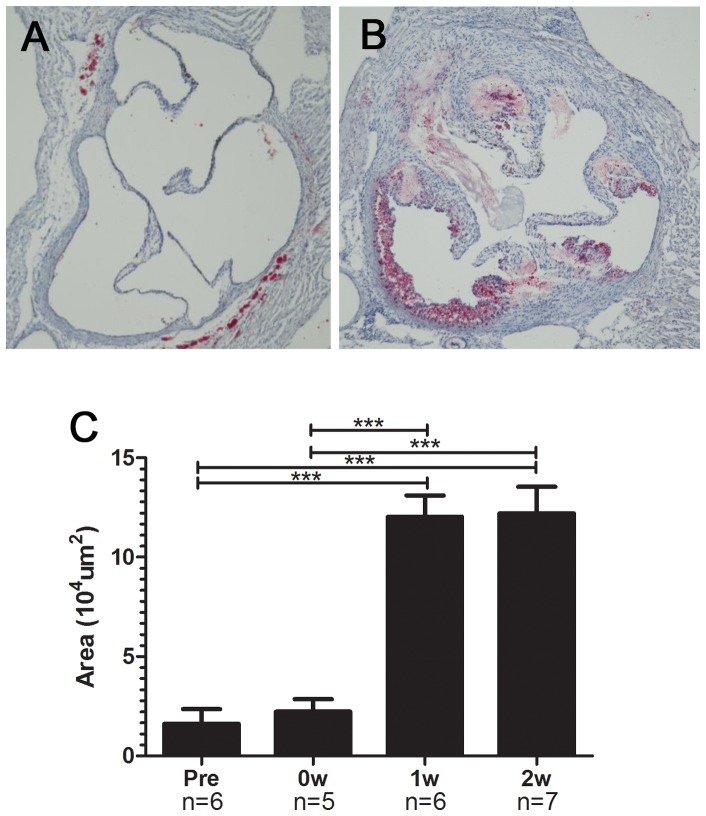
Figure 2. Atherosclerotic lesions in the aorta of modified HypoE mice.
Atherosclerotic lesions at the level of the aortic valve were evaluated by oil red O staining. Representative photographs of specimens taken just before the Paigen diet (A) and 2 weeks after the end of the 7-day Paigen diet intervention (B) are shown. Area of atherosclerotic lesions markedly increased 1 week after the end of the 7-day Paigen diet intervention (C). Pre, 0W, 1W, and 2W represent the same time points as in Figure1. ***P<0.001.

